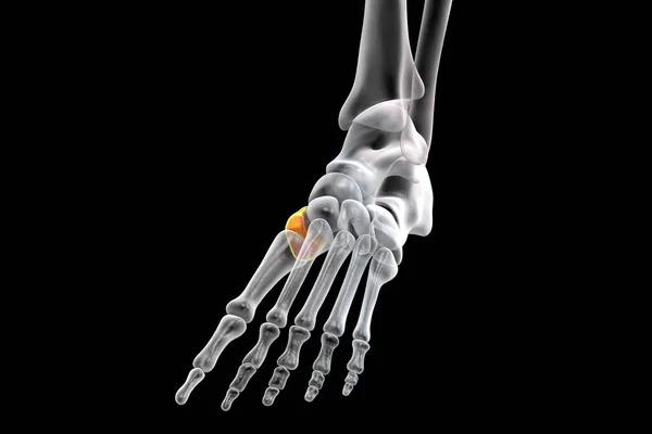
Medial (first) cuneiform bone of the foot, the largest of the cuneiforms. Human foot anatomy. 3D illustration
Esta foto
Medial (first) cuneiform bone of the foot, the largest of the cuneiforms. Human foot anatomy. 3D illustration.
Datos de la Imágen (tiene derechos de autor*)
- Fotografía:
-
Medial (first) cuneiform bone of the foot, the largest of the cuneiforms. Human foot anatomy. 3D illustration
- Autor:
- Ancho original:
-
6000 píxeles.
- Altura original:
-
4000 píxeles.
- Tamaño:
-
24 megapíxeles.
- Categorías:
-
- Palabras Clave:
-
Huesos Aislado póster Primero lado first cuneiform cuerpo frente metatarsiano anatomía falanges Plantar falange cuneiformes ilustración Tobillo Primer plano biología cuboide sanidad diagrama Sistema anatómica proyecciones Cuneiforme medial Fondo negro humano esquelético tarso partes Esqueleto Ciencia Hueso Pie Conjunto Estructura Rayos X Talón astrágalo calcáneo navicular cuneiforme medial Contexto Vista panorámica Médico .
Popularidad
- Vistas:
- 3
- Descargas:
- 0

Fotos similares
Otros temas con fotografías que le puede interesar
- Primero
- Médico
- Vista panorámica
- partes
- Talón
- Esqueleto
- metatarsiano
- falanges
- diagrama
- Ciencia
- falange
- Pie
- cuneiformes
- Tobillo
- póster
- first cuneiform
- Estructura
- esquelético
- medial
- Aislado
- anatomía
- tarso
- Plantar
- Huesos
- Sanidad
- navicular
- Hueso
- anatómica
- frente
- Cuneiforme
- proyecciones
- Contexto
- cuboide
- astrágalo
- Conjunto
- Primer plano
- ilustración
- Rayos X
- Sistema
- cuerpo
- cuneiforme medial
- lado
- biología
- humano
- calcáneo
- Fondo negro
(*) Sitio para adquirir: Link externo para Comprar
Fotografía de Medial (first) cuneiform bone of the foot, the largest of the cuneiforms. Human foot anatomy. 3D illustration, que incluye Medial (first) cuneiform bone of the foot, the largest of the cuneiforms. Human foot anatomy. 3D illustration.
Todas las imágenes por Depositphotos